|
|
BMe Research Grant |
|
Pál Vásárhelyi Doctoral School of Civil Engineering and Earth Sciences
BME Faculty of Civil Engineering, Department of Hydraulic and Water Resources Engineering
Supervisor: Dr. Baranya Sándor
Investigation of the river flow – riverbed interaction with image-based methods
Introducing the research area
The channel formation of the alluvial rivers is influenced by the interaction between the physical properties of the flow (e.g. flow velocity, bed-shear stress, water depth, turbulence) and the riverbed (e.g. composition of the bed material, grain shapes, porosity, stratification). The roughness of the bed surface affects the flow, while the tension, also known as shear stress, originating from the flow itself takes its effects on the sediment making up the riverbed and can incite its movement. Depending on the sediment transport capacity of the river, in some places it washes away, while in other places it builds up its own channel. This complex interaction is studied by the field of hydromorphology. Because of its complex nature, a lot of research is aiming to develop new measurement methods to provide new and never-before-seen results in the topic. Another reason for the high interest is the ever increasing need for economical water management, habitat conservation and climate change. All of these aspects require methodological improvements both in laboratory and field use, because the current procedures are lacking and circumstantial.
Brief introduction of the research place
I carry out my research primarily by using the equipment provided by the Department of Hydraulic and Water Resources Engineering (VVT). The field measurements are done in the upper section of the Hungarian Danube, near to Gönyű settlement. The reasoning behind the choice of the location is the highly various sediment conditions and the practical, engineering issues related to the location. The laboratory measurements are mostly done the in the VVT laboratory. Not long ago, I was able to travel to Norway thanks to a bilateral project and work with the research team of Norwegian University of Science and Technology, Department of Civil and Environmental Engineering (NTNU) after which I came back home with valuable and up-to-date measurement data.
History and context of the research
The channel shape of the natural rivers is constantly evolving. This phenomenon is called morphodynamics. The mechanism has great significance both in human related aspects (e.g. maintaining ship routes, bank infiltration, hydropower) [1,2] and in riverine ecosystems (e.g. fish spawning locations, habitat of macroinvertebrates, dissolved oxygen balance) [3-5]. Despite of its importance, the interaction mechanism is still incites lot of questions. Beside its complexity (but partially also because of it), the traditional sediment measurement methods are not providing satisfactory data neither in quality, nor in quantity. This makes it difficult to fully understand and forecast the interaction in great details. One of the reasons behind it is the conventional measurement methods being time- and energy consuming; the other is their results being point-like information. These are making the measurements even more difficult in the so-called transition zones of the rivers (e.g. Hungarian section of the river Danube) where sediment conditions can change drastically both in space (only in couple hundreds of meters) and time (based on the water regime). If we were to describe this high level of variety, we would need to carry out more measurements, closer to each other, but this is made impossible after a certain point, since the methods are costly. Due to their circumstantial nature, the field measurement campaigns were abandoned in many places, resulting in a disruption of the time series data (sometimes for years). This is mostly true for the bed load measurements. This is coupled with the fact that the conventional bed load samplers are loaded with uncertainties and possible errors [6-10]. Problems are also present in case of bed material (non-moving sediment on the riverbed) and defining their grain size distribution (clay<silt<sand<gravel). For these very reasons, researchers are constantly looking for ways to discover new interrelations of the mechanism and gathering results of better quality.
The research goals, open questions
Nowadays, with the rapid development of informatics and computers, the list of processes that we are able to describe and simulate with numerical models is getting longer. This however requires higher grade of input data and parameters to work properly and indeed proved a more detailed simulation. In the field of hydromorphology however, it is made impossible for the time being due to the above mentioned difficulties and situations. This present study is aiming to fill the gaps and carry out a field measurement methodological improvement, using underwater cameras and so-called image processing methods. This is made possible by the drastic developments in informatics and computer vision. By adapting the proper algorithms and automatizing the analyses, we may be able to cross out the difficulties caused by the time- and energy consuming nature of the traditional methods. Doing this while also gathering direct images of the mechanism, providing several point of view even to visually support of their understanding, further decreasing the uncertainty of the samplings. Last, but not least since the cameras are recording bigger parts of the riverbed, they enable us to analyze bigger areas and not only points.
Methods
In my research I use several image-based methods, depending on the aspect I want to observe and measure. In general, all of the methods handle the images as matrices, where each matrix element is equal to the intensity (grayscale) value of their respected pixels. Then the changing of these values in time and across the picture is analyzed. Another common characteristic of these methods is that they all require a reference object to be visible, which has a known size, so that they can transform the pixel sizes into SI units. The first method I used was the one developed by Buscombe, called “transferable wavelet” method [11] for calculating the grain size distribution of photographed dry sediment. It analyses pixel rows and columns of the pictures and treats the gray-scale values as a sign, processing it with wavelet transformation. I chose this method and adapted it for underwater use. This was made possible by first putting together the right equipment. Finally, it was done by lowering a camera from a motorboat to the riverbed and using scuba lights and a known-sized object, which touched the bed to provide reference for the conversion. Later, this object was replaced by lasers pointing to the riverbed with known distance from each other.

Figure 1 The theory of the wavelet transform by Buscombe. [11]. In the upper left corner the picture of the gravels can be seen with a selected pixel row (black dotted line). The steps of the transformation are shown, resulting in the density function in the bottom right corner. By integrating this, the size distribution is retrieved.

Figure 2 Two examples of underwater images from the very first set of pictures. The reference size was the screw, seen at the bottom of the pictures.
The degree of the change of the riverbed can be well presented by calculating the bed load yield. For this purpose I chose the so-called statistical background method [12,13] which is well-known in surveillance and security systems. I adapted this method and wrote my own code. This method enables the user to separate the moving foreground from the fix background of the video recordings. The first in our case means the bed load, while the latter means the bed material. With this, I was able to estimate the mass of the moving load. For the next step, I worked in collaboration with one of my colleagues and applied a Particle Image Velocimetry (PIV) [14,15] method to define the velocity of the moving particles. After the mass and the velocity were known, the yield could be calculated. After this measurement, I turned my attention towards the Structure-from-Motion [16] procedure, widely used in geoinformatics to create the 3D model of the riverbed, based on the camera recordings. On the one hand, I intended to do this to calculate grain-scale roughness values of the surface; on the other hand I wanted to ensure the possibility of using the generated 3D models in future numerical simulations. This was done by purchasing a commercial geoinformatics software [17] [S.1., S.3.]. Finally, I used the PIV method again, this time however its 2.5D and 3D versions with laser light sources [18-20] and in laboratory environment. In the first case, we measure the 3 velocity components of seeding particles along an enlightened laser sheet projected into the flow, while in the latter case we measure them inside a certain volume. We hypothesize that these particles are behaving exactly the same way as the water particles. I observed the behaviour of the flow over artificial riverbed, built in the laboratory flume.
Results
The first major result of the research was the successful image-based, underwater analysis of the riverbed material on the field. The test measurement results were compared with traditional sampling data in the same spots and for the same water regime. The match between the grain size distributions of the different methods was satisfactory. Also, I managed to handle the theoretical differences originating from the different style of measurements (volume and surface distribution) to a certain level and transformed the original image results into better ones in some cases. The results showed that the applied algorithm can be able to provide the desired fast, satisfyingly precise and map-like data [S.1.,S.3.,S.4.].

Figure 3 Grain size distribution of the riverbed material in a point of the Danube. The traditional method is shown with black, while the image-based original is with blue, the transformed is with yellow. Good matches can be seen in all cases.

Figure 4 Grain size distribution of the riverbed material in a point of the Danube. The traditional method is shown in black, while the image-based original is in blue, the transformed is in yellow. Here, the transformation resulted in a better match.
The next result was the successful, video-based quantification of the bed load. From the underwater recordings, I was able to write my own code and estimate the mass of the moving particles. After coupling this result with the PIV method, the yield value showed good match with the expectations which were based on earlier, traditional sediment measurements. It is also important to mention that this method is compatible with the above mentioned, bed material analyses, since it makes it possible to separate the fix background and the moving foreground of the video, giving the option to analyze them independently [S.3.,S.5.].

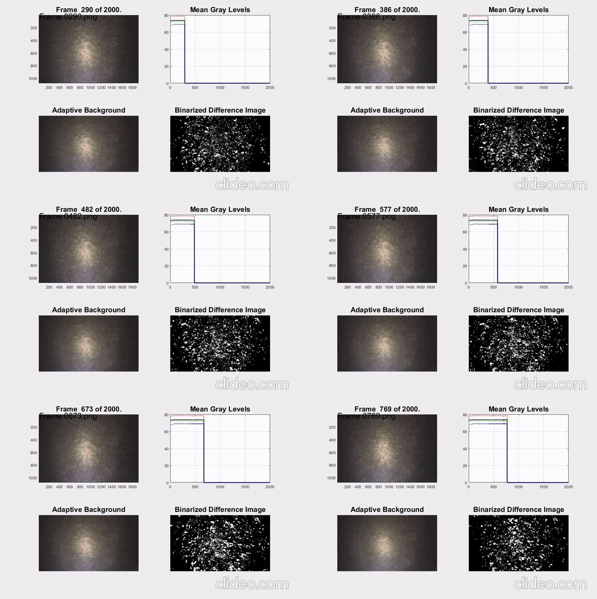
Figure 5 The application of the statistical background method for the video recording. In the upper left corner the original video can be seen. In the bottom right corner the detected moving particles (foreground) are presented with white. In the bottom left corner the fix (background) particles can be seen.

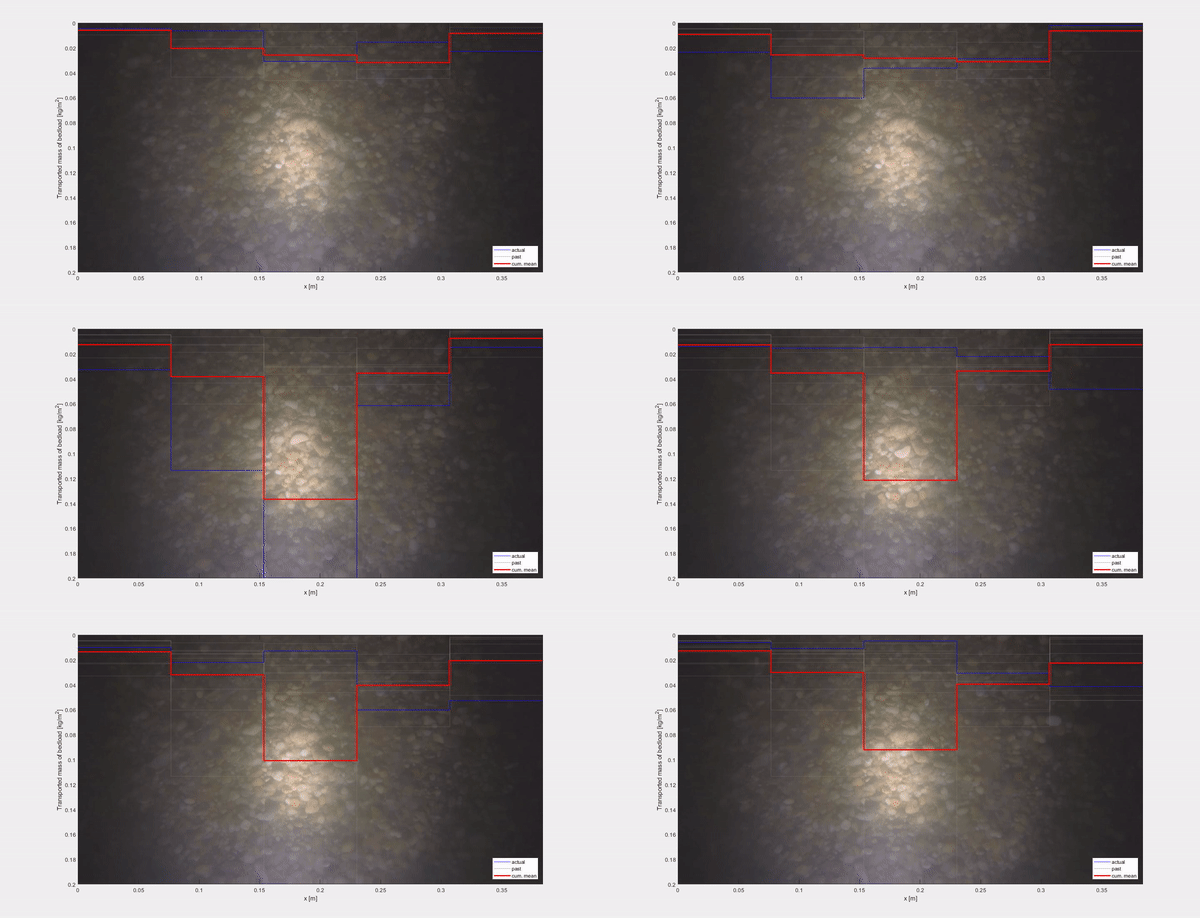
Figure 6 The video was separated into 5 bands to represent the unequal distribution of sediment along the image. On the vertical axis the mass of the moving particles in the given band, in the given instant can be seen in blue, while the cumulated average is shown in red.

Figure 7 The result of the PIV method, the velocity vectors of the recognized patterns.
As a next step, I switched to exploring larger areas with a camera. With the help of the image made during the test measurement, it was possible to map the 3D model of each lane of the riverbed with a resolution of the order of cm, and then to calculate the surface roughness parameter relevant to the bottom sliding stress as a sediment-moving force.


Figure 8 The top two figures show the already concatenated frames of the bed bands, while the bottom two show the 3D bed surface models generated from them. On the left is a more gravelly, rougher, while on the right is a more sandy part.
The values obtained in this way also met the expectations [S.1., S.3.]. In addition to determining roughness, the generated fine-scale 3D terrain can now be incorporated directly into, for example, computer simulation models that can be used to model the movement of water and sediment. At the same time, it is also compatible with the bed material analysis program, as frames can be extracted from the video taken during the scan at any given part of the fixed bed for further analysis. My most recent results are related to fluid dynamics studies in a laboratory glass channel. The applied modern PIV methods allowed a very detailed analysis of the flow over the bed material with different morphological characteristics. With the performed studies, I was able to examine the interaction between turbulence and the bed with rare accuracy, thus contributing to the expansion of the narrow international literature on the topic and to novel results.

Figure 9 A detail of the result of the 2.5D (planar) PIV. The color indicates the magnitude of the resulting velocity of the tracer particles flowing over the bed, which can be assumed to be equal to the velocity of the water itself. The flow is from left to right.

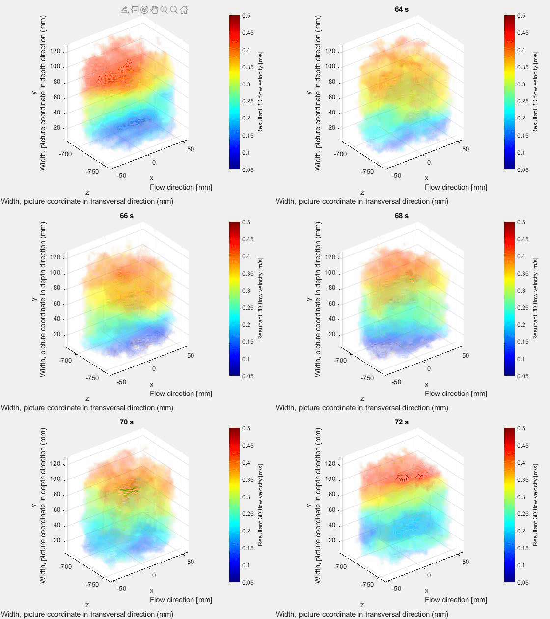
Figure 10 A detail of the result of the 3D (volumetric) PIV. The color indicates the magnitude of the resulting velocity vector. The flow is from right to left.

Figure 11 From a given instantaneous result of the 3D PIV (A) sections can be made either longitudinally (B) or transversely (C). Indicating the direction of the velocity vectors gives a complete picture of the flow structure.
Overall, I have successfully developed and validated a new image-based field measurement method in the field of hydromorphology. I explored the applicability limits of each procedure, and then based on them I also identified possible directions for further development. In the course of my research, I further developed and adapted image processing methods from other disciplines that can be accessed and interconnected. All methods are compatible with bed material analysis, but, for example, models of bed bands made with the Structure-from-Motion method can even be transferred to the laboratory using 3D printing for PIV testing. This degree of interconnection of methods also demonstrates that their application can be an innovation to provide a more accurate understanding of the measurement and interaction desired by the profession.

Figure 12 An example of combining the methods presented. A) A 3D model of the bed surface produced by bed structure imaging using the Structure-from-Motion method. Roughness calculations were performed along the red line shown in the image (intersecting the model). B) Linear section of the bed surface and changes in the calculated roughness values. C) A photograph of the riverbed taken at one point in the camera, with individual particles identified and measured in blue by the image processing algorithm. D) Laboratory result on the velocity field above the bed (dark blue) in the so-called Particle Image Using Velocimetry image processing procedure.
Expected impact and further research
The series of measurement technology developments carried out during the research, making them practical and accessible, is intended to fill the gaps in hydromorphological studies so far, producing novel and unique results both at the domestic and international level. The new information available in this way can help describe the interaction between river flow and the riverbed at a higher level than before, and to find new connections. In the future, I plan a detailed analysis of the results of field and glass channel laboratory measurements. Once this is done, I will perform numerical modeling studies on sediment motion in the flow structures and conditions known in detail in the laboratory, taking advantage of the fact that the “ground state” (without moving sediment) was assessed very well with the new methods. If the laboratory computer models confirm the correlations revealed, their application to field conditions will be the next step. The video-based procedure developed during the research, which explores the migration of rolling sediment moving near the bedrock, attracted the interest of the Norwegian NTNU University, and on their initiative we developed the foundations of a Hungarian-Norwegian-German joint research project. The first part of the research results presented here was published in a domestic and a highly quoted international journal (WATER), proving the importance and topicality of the topic. I have also presented my results at international conferences, and as a result, a source code has been exchanged with one of the renowned professors at ETH Zurich, Switzerland [21].
Publications, references, links
List of corresponding own publications
S.1. Ermilov, A. A., Baranya, S., & Török, G. T. (2020). Image-Based Bed Material Mapping of a Large River. WATER, 12(3). http://doi.org/10.3390/w12030916
S.2. Ermilov, A. A. (2020). „Mossa a Duna? Hiszem, ha látom.” - Innovatív mérési módszerek fejlesztése folyó hidromorfológiában. ÉLET ÉS TUDOMÁNY, Vol. LXXV. (No. 31), 969–971.
S.3. Ermilov, A. A., Baranya, S., Fleit, G., & Török, G. T. (2020). Képalapú módszerek fejlesztése folyók morfodinamikai vizsgálatához. HIDROLÓGIAI KÖZLÖNY.
S.4. Ermilov, A. A., Baranya, S., & Török, G. T. (2019). IMAGE BASED BED MATERIAL MAPPING OF A LARGE RIVER. In Proceedings of the 2nd International Symposium and Exhibition on Hydro-Environment Sensors and Software (pp. 105–112).
S.5. Ermilov, A. A., Fleit, G., Zsugyel, M., Baranya, S., & Török, G. T. (2019). Video based bedload transport analysis in gravel bed rivers. In Geophysical Research Abstracts (Vol. 21).
S.6. Benkő, G., Baranya, S., Török, G. T., Molnár, B., & Ermilov, A. A. (2019). Analysis of river bed material composition with Deep Learning based on drone video footages. In Geophysical Research Abstracts (Vol. 21).
S.7. Ermilov, A. A., Baranya, S., & Rüther, N. (2018). Numerical simulation of sediment flushing in reservoirs with TELEMAC. GEOPHYSICAL RESEARCH ABSTRACTS, 20 (EGU2018-12355), 12355 (2018).
Table of links
Department of Hydraulic and Water Resources Engineering
Norwegian University of Science and Technology, Department of Civil and Environmental Engineering
volume and surface distribution
List of references
1. Healey, K. M., Cox, A. L., Hanes, D. M., Chambers, L. G. (2015). State of the practice of sediment management in reservoirs: Minimizing sedimentation and removing deposits. Proceedings of the 5th Federal Interagency Hydrologic Modeling Conference and the 10th Federal Interagency Sedimentation Conference, April 19-23, 2015.
2. Rákóczi, L. (1979). Mederanyag-minták információtartalma és hasznosítása a folyószabályozásban. Magyar Hidrológiai Társaság Országos vándorgyűlésének kiadványa, Keszthely, Hungary
3. Buendia, C., Gibbins, C. N., Vericat, D., Batalla, R. J., Douglas, A. (2013): Detecting the structural and functional impacts of fine sediment on stream invertebrates. Ecol. Indic. 25: 184-196. https://doi.org/10.1016/j.ecolind.2012.09.027.
4. Descloux, S., Datry, T., Marmonier, P. (2013): Benthic and hyporheic invertebrate assemblages along a gradient of increasing streambed colmation by fine sediment. Aquati Sci. 75 (4): 493-507. https://doi.org/10.1007/s00027-013-0295-6
5. Sear, D. S., Frostick, L. B., Rollingson, G., Lisle, T. E. (2008): The Significance and Mechanics of Fine-sediment Infiltration and Accumulation in Gravel Spawning Beds. American Fisheries Society Symposium 65.
6. Ehrenberger, R. (1931). Direkte Geschiebemessungen an der Donau bei Wien und deren bisherige Ergebnisse. Die Wasserwirtschaft, 34: 581–589.
7. Emmett, W.W. (1980). A field calibration of the sediment-trapping characteristics of the Helley-Smith bedload sampler. USGS Professional Paper, 1139, U.S. Govt. Print. Off.
8. Carey, W.P. (1985). Variability in measured bedload-transport rates. Water Resources Bulletin 21 (1), 39–48.
9. Vericat, D., Church, M., Batalla, R.J. (2006). Bed load bias: comparison of measurements obtained using two (76 and 152 mm) Helley–Smith samplers in a gravel-bed river. Water Resour. Res. W01402. http://dx.doi.org/10.1029/2005WR004025
10. Camenen, B., Jaballah, M., Geay, T., Belleudy, P., Laronne, J. B., and Laskowski, J. P. (2012). Tentative measurements of bedload transport in an energetic alpine gravel bed river. River Flow 2012, Taylor & Francis Group, London, 379–386, 2012.
11. Buscombe, D. (2013). Transferable wavelet method for grain-size distribution from images of sediment surfaces and thin sections, and other natural granular patterns. SEDIMENTOLOGY (2013) 60, 1709-1732. doi: 10.1111/sed.12049.
12. Bouwmans, T. (2010). Statistical Background Modelling for Foreground Detection: A Survey. Handbook of Pattern Recognition and Computer Vision, pp. 181–199. doi: 10.1142/9789814273398_0008.
13. Jeeva, S., Sivabalakrishnan, M. (2015). Survey on background modeling and foreground detection for real time video surveillance. Procedia Computer Science 50 (2015) 566 – 571. ISBCC’15. https://doi.org/10.1016/j.procs.2015.04.085
14. Adrian, R. J. (1991). Particle-Imaging Techniques for Experimental Fluid Mechanics. Annu. Rev. Fluid Mech. 23 (1991) 261–304.
15. Fleit, G., Baranya, S. (2019). An improved particle image velocimetry method for efficient flow analyses. Flow Measurement and Instrumentation Vol. 69, October 2019, 101619. https://doi.org/10.1016/j.flowmeasinst.2019.101619
16. Westoby, M. J., Brasington, J., Glasser, N. F., Hambrey, M. J., Reynolds, J. M. (2012). ‘Structure-from-Motion’ photogrammetry: A low-cost, effective tool for geoscience applications. Geomorphology, Volume 179, 15 December 2012, pp. 300–314. https://doi.org/10.1016/j.geomorph.2012.08.021
17. Agisoft LLC (2019). Agisoft Metashape User Manual: Professional Edition, Version 1.5. 2019, https://www.agisoft.com/downloads/user-manuals/
18. Adrian, R. J. (1991). Particle-Imaging Techniques for Experimental Fluid Mechanics. Annu. Rev. Fluid Mech. 23 (1991) 261–304.
19. Acevedo-Ávila, R., González-Mendoza, M., Garcia-Garcia, A. (2007). A Statistical Background Modeling Algorithm for Real-Time Pixel Classification. ISSN 2007-9737. Computación y Sistemas, Vol. 22, No. 3, 2018, pp. 917–927.doi: 10.13053/CyS-22-3-2554
20. Unsworth, A. Ch. (2015). Particle Imaging Velocimetry. Geomorphological Techniques, Chap. 3, Sec. 3.4 (2015)
21. Detert, M., Weitbrecht, V. (2012). Automatic object detection to analyze the geometry of gravel grains - A free stand-alone tool. River Flow 2012 Conference – Murillo (Ed.).
