|
|
BMe Research Grant |
|
George A. Olah Doctoral School of Chemistry and Chemical Technology
MTA Wigner Research Centre for Physics
Supervisor: Dr. Gali Ádám
Silicon Carbide Nanoclasters for Bioimaging and Quantum computing
Introducing the research area
Silicon Carbide1 (SiC) is an excellent material known as semiconductor2 that may substitute silicon in high power electronics [1] and as ceramic3 with the hardness of diamond [2]. SiC shows new properties in nano size where quantum confinement4 takes place. The surface of SiC nanocrystal acts like an organic compound while the core crystal becomes an excellent luminescence source opening the door for new generations of luminescent dyes5. I synthesize these nanocrystals and study their chemical and physical properties. My mission is to understand the physics behind these nanoclusters and make the best dyes possible for in vivo bioimaging by changing the physical and chemical properties of the material.
Brief introduction of the research place
My PhD work has been carried out as a member of the group of Prof. Adam Gali at Wigner Research Centre6. Wigner RCP was founded by uniting two former research institutes, the Research Institute for Particle and Nuclear Physics, and the Research Institute for Solid State Physics and Optics of the H.A.S. Our group’s7 research focuses on theoretical and experimental characterization of silicon carbide based nanoclusters.
History and context of the research
Visual analysis of biological systems is an integral avenue of basic and applied biological research. Fluorescence microscopy techniques are often used for studying intracellular mechanisms [3]. The importance of this method is clearly demonstrated by the 2014 Nobel Prize8 in chemistry that has been awarded for the ultrahigh resolution microscopy9 [4], but fluorescence dyes have been used to label cancer cells10 during surgery too [5a,b].
There are some groups who already synthesized SiC nanocrystals [6a,b] and studied their physiological properties [7], but the first methods were unable to provide SiC nanoclusters with molecular size and narrow size distribution ‒ both properties are essential for biological applications and to understand their optical properties.
The research goal, open questions
My research goal is to synthesize SiC nanocrystals that are suitable for in-vivo11 and in-vitro bioimaging. To this end, nanocrystals should be biocompatible, sufficiently small for clearance [8] after the treatment, and applicable in aqueous environment. While most dyes are toxic [9], the biocompatibility of SiC is known for a long time [10]. The first task was to find proper synthesis method that is capable of producing SiC nanocrystals in large quantities with narrow size distribution. The origin of luminescence has to be clarified too. A further aim is to make the emission size-independent through the inclusion of luminescence centers12. Certain luminescence centers in SiC act as single photon source which makes our nanocrystals suitable for quantum information processing as well.

Figure 1: Vacancies in SiC
Methods
I used several experimental methods. For producing SiC nanocrystals we have to use top-down methods and bulk SiC. Development of bulk SiC synthesis is crucial for luminescence centers too. We used volume synthesis13 method, a fast reaction between silicon and carbon [11].
To prepare SiC in the 1‒4 nm range, another chemical reaction, namely wet chemical etching [6b] was necessary. SiC is highly resistant material, but with a mixture of hydrofluoric acid and nitric acid at 100 °C the reaction takes place.

Figure 2: Synthesis of SiC nanocrystals
For further measurements, SiC nanocrystals were dispersed in a suitable solvent. Optical properties of the colloid sol14 were determined by fluorescence spectrometer15 and we applied infrared spectroscopy16 to study their surface chemistry

Figure 3: Infrared spectrum of SiC nanocrystals.
Results
I arrived at Gali’s group at a very early stage of the experimental research. My first task was the elaboration of synthesis method for SiC nanocrystals at our Institute. I developed a synthesis route based on literature by applying closed reaction chamber, similar to the one used for hydrothermal synthesis [B1, B2]. My development enhances production safety, yield, and quality. The size of our nanocrystals is about 1‒3 nm, a size distribution very rare at top-down methods17.
I developed the synthesis of bulk SiC, as well by applying changes to the existing furnace system and to the reaction route. Thanks to the applied developments, we are now able to synthesize SiC nanocrystals with better quality [B3], and can synthesize SiC nanocrystals of about 100 nm size containing red single photon centers [B4] with high density18.
In the case of the smallest 1‒3 nm nanocrystals, the luminescence is very complex mechanism where quantum confinement and surface chemistry jointly determine the emission. The energy of emitted photons is highly influenced by the oxygen containing surface groups which relation was proven both experimentally and theoretically [B5, B6]. I also studied the effect of different surface groups (carboxyl, carbonyl, hydroxyl, etc.) by making different surface terminated SiC nanocrystals. Using time dependent fluorescence spectroscopy19 we found an increase in the emission wavelength with increasing reduction degree of the surface. We also found another emission center which is connected to the Si-O bonds on the surface.

Figure 4: Emission vs. surface at SiC nanocrystals.
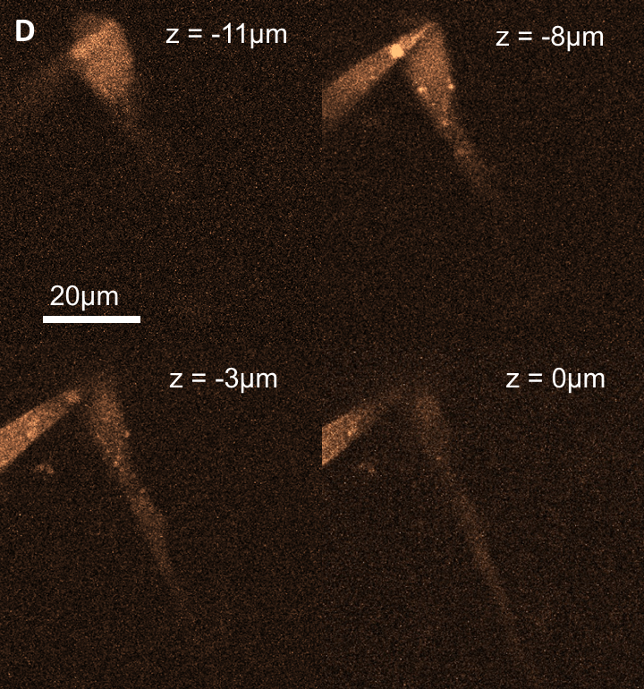
We took a small step toward the application of the method by measuring the toxicity of the nanocrystals and using SiC as fluorescence probe for two-photon imaging [B7].

Figure 5: SiC nanocrystal labeled neuron cell under two-photon microscope.
Expected impact and further research
We have already proved the applicability of SiC nanocrystals in biological systems but the real break-through would be the synthesis of SiC nanoclusters with red-infrared emission. To this end, we have to synthesize SiC nanocrystals containing red emission centers but we have to optimize the surface of the nanoparticles too. Currently we are able to prepare SiC nanocrystals with 620 nm emission. My next step is to analyze this emission center and to further optimize the synthesis method. Understanding the connection between surface chemistry and luminescence, and the identification of the new emission center would allow us creating a new, SiC-based dye and nanosensor family with low toxicity and clearance of SiC, which could open new opportunities in biology and quantum computing.
Publications, references, links
Publications related to this topic (IF: impact factor, C: independent citations)
Number of publications: 8, total impact factor: 40.145, Independent citations: 61
[B1]. Beke D, Szekrenyes Zs, Balogh I, Veres M, Fazakas E, Varga L.K, Kamaras K, Czigany Zs, Gali A., Characterization of luminescent silicon carbide nanocrystals prepared by reactive bonding and subsequent wet chemical etching, Applied Physics Letters, 99(21) 213108. (2011).
IF: 3,142; C: 13
[B2] Beke D, Szekrényes Z, Balogh I, Czigány Z, Kamarás K, Gali A., Preparation of small silicon carbide quantum dots by wet chemical etching, Journal of Materials Research, 28(1), 44 (2013).
IF: 1,673; C: 9
[B3] Szekrényes Zs., Somogyi B., Beke D., Károlyházi Gy., Balogh I., Kamarás K., Gali A., Chemical Transformation of Carboxyl Groups on the Surface of Silicon Carbide Quantum Dots, Journal of Physical Chemistry C – Nanomaterials and Interfaces, 118(34), 19995, (2014).
IF: 4,509; C: 3
[B4] Old B1Castelletto S., Johnson B, Zachreson C., Beke D., Balogh I., Ohshima T. Aharonovich I., Gali A., Room Temperature Quantum Emission from Cubic Silicon Carbide Nanoparticles, ACS Nano, 8(8), 7938, (2014).
IF: 13,334; C: 12
[B5] Beke D., Szekrényes Zs., Czigány Zs., Kamarás K., Gali A., Dominant Luminescence is not Due to Quantum Confinement in Molecular Sized Silicon Carbide Nanocrystals, Nanoscale 7:(25) 10982. (2015).
IF: 7,760; C: 7
[B6] Beke D., Jánosi TZ, Somogyi B, Major D. Á, Szekrényes Zs , Erostyák J , Kamarás K., Gali A., Identification of Luminescence Centers in Molecular-Sized Silicon Carbide Nanocrystals, JPC. C, 120:(1) 685. (2016).
IF: 4,509; C: 6
[B7] Beke D., Szekrényes Zs., Pálfi D., Róna G., Balogh I., Maák P.A., Katona G., Czigány Zs., Kamarás K., Rózsa B., Buday L., Vértessy B., Gali A., Silicon carbide quantum dots for bioimaging, Journal of Materials Research, 10(28), 205 (2013)
IF: 1,673; C: 9
Other publications in this topic
Dravecz G, Bencs L, Beke D, Gali A., Determination of silicon and aluminum in silicon carbide nanocrystals by high-resolution continuum source graphite furnace atomic absorption spectrometry, TALANTA 147., 271. (2016)
Beke Dávid: Kvantumpöttyök – biológiai képalkotás, Természet Világa, 145(9), 396 (2014)
Beke D, Szekrényes Zs, Róna G, Pálfi D, Vértessy B, Rózsa B, Kamarás K, Gali Á Silicon Carbide Quantum Dots As A Non-Toxic Probe For Bioimaging: Synthesis And Characterization In: Szilárd Szélpál (ed.) I. Innovation in Science - Doctoral Student Conference 2014: eBook of Abstracts. 207 p. (Doktoranduszok Országos Szövetsége, Biológiai és Kémiai Tudományok Osztálya) Szeged: Magyar Kémikusok Egyesülete, 2014. pp. 30-31. (ISBN:978-963-9970-52-6)
Jánosi TZ, Beke D, Szekrényes Zs, Kamarás K, Gali A, Erostyák J Szilicium-karbid kvantum dotok fluoreszkáló centrumainak szétválasztása időemissziós mátrix analízisével In: Ádám P, Almási G (ed.) Kvantumelektronika 2014: VII. Szimpózium a hazai kvantumelektronikai kutatások eredményeiről. P09. 2 p. (ISBN:978-963-642-697-2)
Beke D, Szekrényes Zs, Kamarás K, Gali Á Lumineszcens szilíciumkarbid kvantumpöttyök előállítása és jellemzése In: Keresztes Gábor (ed.) Tavaszi Szél, 2013: Spring wind, 2013. 659 p. Budapest: Doktoranduszok Országos Szövetsége, 2013. pp. 62-71. (ISBN:978-963-89560-2-6)
Links
1. http://en.wikipedia.org/wiki/Silicon_carbide
2. http://spinoff.nasa.gov/Spinoff2008/ip_2.html
3. http://www.reade.com/Particle_Briefings/mohs_hardness_abrasive_grit.html
4. https://youtu.be/9RmBRoHctDI
5. https://youtu.be/gSGq8gOLXwY
7. http://wiki.kfki.hu/nano/Semiconductor_Nanostructures_%E2%80%9CLend%C3%BClet%E2%80%9D_Research_Group
8. http://www.nobelprize.org/nobel_prizes/chemistry/laureates/2014/
9. http://en.wikipedia.org/wiki/Super-resolution_microscopy
10. http://blogs.discovermagazine.com/d-brief/2015/04/08/fluorescent-dye-tumors/#.VXgmUs-qpBc
11. http://en.wikipedia.org/wiki/In_vivo
12. http://wiki.kfki.hu/nano/Fluorescent_semiconductor_nanocrystals_for_biological_imaging
13. https://youtu.be/eUs2__t--3s
14. http://en.wikipedia.org/wiki/Colloid
15. http://en.wikipedia.org/wiki/Fluorescence_spectroscopy
16. http://en.wikipedia.org/wiki/Infrared_spectroscopy
18. http://www.prolibraries.com/mrs/?select=new_speaker&speakerID=69948&view=simple&conferenceID=12
19. http://en.wikipedia.org/wiki/Time-resolved_spectroscopy
References
[1]: J.B. Casady, R.W. Johnson, Status of silicon carbide (SiC) as a wide-bandgap semiconductor for high-temperature applications: A review, Solid-State Electronics 39(10), 1409–1422 (1996)
[2]: R. M. Laine , F. Babonneau, Preceramic polymer routes to silicon carbide, Chem. Mater, 5(3), 260–279 (1993)
[3]: Z. Yao, R. Carballido-López, Fluorescence Imaging for Bacterial Cell Biology: From Localization to Dynamics, From Ensembles to Single Molecules, Annual Review of Microbiology 68, 459-476, (2014)
[4]: Z. Liu, L. D. Lavis, E. Betzig, Imaging Live-Cell Dynamics and Structure at the Single-Molecule Level, Molecular Cell 58(4), 644–659 (2015)
[5a]: C. Chi, Y. Du, J. Ye, D. Kou, J. Qiu, J. Wang, J. Tian, X. Chen, Intraoperative imaging-guided cancer surgery: from current fluorescence molecular imaging methods to future multi-modality imaging technology, Theranostics. 4(11), 1072-84, (2014)
[5b]: A. L. Vahrmeijer, M. Hutteman, J. R. van der Vorst, C. J. H. van de Velde, J. V. Frangioni, Image-guided cancer surgery using near-infrared fluorescence, Nature Reviews Clinical Oncology 10, 507–518 (2013)
[6a]: V. Buschmann, S. Klein, H. Fueß, H. J. Hahn, HREM study of 3C–SiC nanoparticles: influence of growth conditions on crystalline quality, Crystal Growth, 193, 335, (1998)
[6b]: X.LWu, J. Y. Fan, T. Qiu, X. Yang, G. G. Siu, P. K. Chu, Experimental Evidence for the Quantum Confinement Effect in 3C-SiC Nanocrystallites. Phys Rev Lett 94, 026102 (2005)
[7]: J. Botsoa, V. Lysenko, A. Géloen, O. Marty J. M. Bluet, G. Guillot . Application of 3C-SiC quantum dots for living cell imaging. Appl. Phys. Lett. 92, 173902 (2008).
[8]: H. S. Choi, W. Liu, P. Misra, E. Tanaka, J. P. Zimmer, B. I. Ipe, M. G. Bawendi, J. V. Frangioni, Renal Clearance of Nanoparticles, Nat Biotechnol. 25(10), 1165–1170, (2007)
[9]: A. Valizadeh, H. Mikaeili, M. Samiei, S. M. Farkhani, N. Zarghami, M. Kouhi, A. Akbarzadeh, S. Davaran, Quantum dots: synthesis, bioapplications, and toxicity, Nanoscale Research Letters, 7, 480, (2012)
[10]: C. Coletti, M.J. Jaroszeski, A. Pallaoro, A.M. Hoff, S. Iannotta, S.E. Saddow, Biocompatibility and wettability of crystalline SiC and Si surfaces, Conf Proc IEEE Eng Med Biol Soc.2007 5850, (2007)
[11]: Alexander S. Mukasyan (2011). Combustion Synthesis of Silicon Carbide, Properties and Applications of Silicon Carbide, Prof. Rosario Gerhardt (Ed.), ISBN: 978-953-307-201-2, InTech,
